3. INSTRUMENTS
The details of experimental technique for x-ray and neutron structure analysis involving small-angle scattering instrumentation is reviewed by Glatter and Kratky (Glatter, 1982) and Schelten and Hendricks (Schelten, 1978). Here I deal only with some examples of SAXS and SANS instruments for getting 'images' of such the experimental technique.
3.1) SAXS spectrometer at laboratory
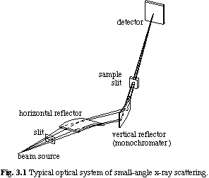
A standard setup of SAXS spectrometer is constructed with x-ray source, optical system, and a counter. All the elements have much variations depending on purpose (objects for experiments), Q-range, and angular resolution. In the small-angle scattering, a position sensitive counter covering a large area at small-Q region is frequently used in order to measure the scattering neighboring the direct beam. In such cases, it is usual to use point focus x-ray source in order to avoid the labor of the slit compensation in handling the data. In addition, it is necessary that a sample is measured in the transmission configuration like the "Laue case" in the usual crystal diffraction. (The "Bragg case" in the crystallography may correspond to the reflectometry.) Typical setup of a small-angle x-ray scattering apparatus is shown in Fig. 3.1. A beam source is a Cu-target rotating anode x-ray generator, a bent quartz mirror and a bent graphite crystal being used to focus and monochromatize the incident beam. A sample is placed just behind the monochrometor, and an area detector (a position sensitive proportional counter or an imaging plate) is placed at a few tens cm behind the sample. A vacuum tube is inserted between the sample and the detector in order to avoid a strong small-angle scattering from the air. A typical beam size is 1 mm x1 mm at the sample position, and a typical exposure time is a few hours.

3.2) SAXS spectrometer at synchrotron radiation x-ray source
Because small-angle scattering from most of objects, especially from solutions, are too weak to investigate at laboratory-level x-ray source, it is efficient to use synchrotron radiation x-ray source. Figure 3.2 shows an arrangement of the SAXS spectrometer of BL-15A at Photon Factory, High Energy Accelerator Research Organization (KEK), Tsukuba, Japan as a typical example. X-ray beam is taken out from bending magnet, monochromatized and focused on the counter position by a set of bent quartz mirror and bent silicon single crystal, and the scattered beam is detected by three types of area detectors; one-dimensional position sensitive proportional counter, imaging plate, or CCD camera. A counter position is fixed at 4.6 m apart from the monochrometer, and a sample is placed between a few tens cm and 3 m before the counter. Typical beam size at the sample position is 1.5 mm high and 2.6 mm wide. Details of this spectrometer was shown by Amemiya et al. (Amemiya, 1983). Although the incident beam intensity of this spectrometer is not very strong comparing with other newer synchrotron sources, an expected x-ray intensity of 1011 - 1012 photons/s is several orders of magnitude more than a SAXS spectrometer at laboratory. Therefore, this type of spectrometer is suitable to investigate dynamical structure of objects within a time scale of sub-milli seconds.
3.3) SANS spectrometer at research reactor
Recently, majority of objects for SAS investigation are so-called "soft materials", which include polymers, colloids, biological systems, liquid crystals, complex fluids, and so on. These systems mainly constructed with hydrogen, carbon, oxygen and other light atoms. Therefore, neutron is much more useful than x-ray in order to investigate a structure of these systems.
Figure 3.3 shows a configuration of SANS-U spectrometer at JRR-3M of Japan Atomic Energy Research Institute (JAERI), Tokai, Japan (Ito, 1995) as a typical example. Incident neutron beam is taken from a cold neutron source of the research reactor, and monochromatized by a mechanical velocity selector. Typical wavelength is 7 Å and its resolution is about 10 %, and a position of a two-dimensional proportional counter from the sample position can be varied from 1 m to 12 m depending on the objective Q-range. Typical beam size at the sample position is about 10 mmf, and an exposure time is from 10 minutes to a few hours.
3.4) SANS spectrometer at spallation neutron source
Another type of SANS spectrometers are installed at spallation neutron sources. It uses a pulsed neutron with the wide wavelength range, and the time-of-flight analysis should be performed to get an information of a Q-value of scattered neutrons with information of a scattering angle. An advantage of spectrometers at spallation sources against ones at reactor is that wide Q-range is observable simultaneously. In Fig. 3.4, schematic diagram of SWAN spectrometer at KENS of KEK is shown (Otomo, 1999). Detectors are placed not only at the small angle region but also at high angle. Therefore, the observable Q-range widely spreads from 0.005 Å-1 to 12 Å-1. The Q resolution (DQ/Q) at small-angle scattering region is about 30 %, and the beam size at the sample is about 2 cm square. Typical exposure time is several hours. Note that the beam source of KEK is the first generation one and its intensity is one or two orders of magnitude less than the other newer sources. Another paper should be referred (for example, Tyiyagarajan, 1997) in order to compare a performance of TOF-SANS with other SAS spectrometers.